Home » Without Label » Anatomy Of The Upper Chest Area - Diagram Chest Muscles Women - 508 best Chest images on ... : Thoracic cavity, also called chest cavity, the second largest hollow space of the body.it is enclosed by the ribs, the vertebral column, and the sternum, or breastbone, and is separated from the abdominal cavity (the body's largest hollow space) by a muscular and membranous partition, the diaphragm.it contains the lungs, the middle and lower airways—the tracheobronchial tree—the heart.
Anatomy Of The Upper Chest Area - Diagram Chest Muscles Women - 508 best Chest images on ... : Thoracic cavity, also called chest cavity, the second largest hollow space of the body.it is enclosed by the ribs, the vertebral column, and the sternum, or breastbone, and is separated from the abdominal cavity (the body's largest hollow space) by a muscular and membranous partition, the diaphragm.it contains the lungs, the middle and lower airways—the tracheobronchial tree—the heart.
Anatomy Of The Upper Chest Area - Diagram Chest Muscles Women - 508 best Chest images on ... : Thoracic cavity, also called chest cavity, the second largest hollow space of the body.it is enclosed by the ribs, the vertebral column, and the sternum, or breastbone, and is separated from the abdominal cavity (the body's largest hollow space) by a muscular and membranous partition, the diaphragm.it contains the lungs, the middle and lower airways—the tracheobronchial tree—the heart.. It is mostly protected and supported by the rib cage, spine, and shoulder girdle. It runs across the front of your neck and behind the clavicle (collarbone) to supply blood to the muscles, skin, and bones in your chest and shoulder. Anatomy of the chest and shoulder, anatomy of the chest organs, anatomy of the chest wall, anatomy of the chest wall and pleura, anatomy of upper chest area, human. In humans and other hominids, the thorax is the chest region of the body between the neck and the abdomen, along with its internal organs and other contents. The epidermis is the outermost layer that provides a protective, waterproof seal over the body.
Browny/reddy colour only appears when the body's immune system begins to decay with the digestive organs. Anatomy of the chest and the lungs: System respiratory respiratory organs of human body digestive and respiratory system medical chest internal structure of human body medicine body lungs biology intestines stomach anatomy torso human internal. The ribs and sternum make up what is called the 'ribcage.' the ribcage protects the lungs, blood vessels, and heart,. Anatomy as mentioned above, the trapezius muscle is divided into 3 areas:
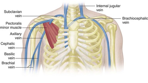
Venous Sonography of the Upper Extremities and Thoracic ... from radiologykey.com In the front, the neck extends from the bottom part of the mandible (lower jaw bone) to the bones of the upper chest and shoulders (including the sternum and collar bones). The muscles of the chest and upper back occupy the thoracic region of the body inferior to the neck and superior to the abdominal region and include the muscles of the shoulders. It contains four muscles that exert a force on the upper limb: It starts from the pharynx and extends to the upper end of the esophagus. Chest defends and protects the breathe and breathing system. Webmd's colon anatomy page provides a detailed image and definition of the colon. Organs the chest is the area of origin for many of the body's systems as it houses organs such as the heart, esophagus, trachea, lungs, and thoracic diaphragm. Anatomy of the chest and shoulder, anatomy of the chest organs, anatomy of the chest wall, anatomy of the chest wall and pleura, anatomy of upper chest area, human.
The suprascapular artery is a branch of the thyrocervical trunk, which emerges from the first part of the subclavian artery.
The pec major itself is comprised of two heads, which jointly attach to your upper arm. See chest anatomy stock video clips. Upper back pain and chest pain can occur together. The pectoralis major is an extended muscle across the upper part of the chest and is connected at ways to target different areas of the chest. Anatomy of the upper chest area. The nervous system of the thorax is a vital part of the nervous system as a whole, as it includes the spinal cord, peripheral nerves, and autonomic ganglia that communicate with and control many vital organs. The circulatory system does most of. It describes the theatre of events. 1 the division into the separate, distinct parts of this muscle is about functionality. In other words, each area does something different. An anatomical guide to training : Learn about its function, location, and conditions that affect the colon. The epidermis is the outermost layer that provides a protective, waterproof seal over the body.
Each one spans half of the upper chest, and has attachment points on the sternum (breastbone), ribs, clavicle (collarbone), and humerus (long bone of your upper arm). The neck is a complex anatomic region between the head and the body. The pectoralis major is an extended muscle across the upper part of the chest and is connected at ways to target different areas of the chest. In the front, the neck extends from the bottom part of the mandible (lower jaw bone) to the bones of the upper chest and shoulders (including the sternum and collar bones). The major muscle in the chest is the pectoralis major.
Chest Muscle Anatomy Diagram - Pectoral Muscles Area ... from media.sciencephoto.com In other words, each area does something different. Huge collection, amazing choice, 100+ million high quality, affordable rf and rm images. Your torso consists of two parts — the chest and the abdomen. When the throat is hurt, the chest must bear some pain or the body is in other major diseases. Nerves of the chest and upper back. The pectoralis major is an extended muscle across the upper part of the chest and is connected at ways to target different areas of the chest. Anatomy of the upper chest area : The circulatory system does most of.
At the level of the pelvic bones, the abdomen.
Anatomy as mentioned above, the trapezius muscle is divided into 3 areas: Anatomy of the chest and the lungs: The chest contains your heart and lungs; The abdomen (commonly called the belly) is the body space between the thorax (chest) and pelvis. The back of the neck is mostly comprised of muscles, as well as the spine. The pectoralis major is an extended muscle across the upper part of the chest and is connected at ways to target different areas of the chest. Disorder of the esophagus makes swallowing difficult and sometimes painful. It is the level 2 symptom to this problem. Three dimensional view of the female reproductive system, full frontal view. Learn about its function, location, and conditions that affect the colon. 1 the division into the separate, distinct parts of this muscle is about functionality. It runs across the front of your neck and behind the clavicle (collarbone) to supply blood to the muscles, skin, and bones in your chest and shoulder. The muscles of the chest and upper back occupy the thoracic region of the body inferior to the neck and superior to the abdominal region and include the muscles of the shoulders.
Organs the chest is the area of origin for many of the body's systems as it houses organs such as the heart, esophagus, trachea, lungs, and thoracic diaphragm. The pectoral region is located on the anterior chest wall. The sternum, or breastbone, is a flat bone at the front center of the chest. Understanding the basics of throat anatomy with diagram and pictures. The mammary bud grows downward into the dermis and starts branching to the secondary bud around the twelfth week.
What are the problems that can be caused by undeveloped ... from qph.fs.quoracdn.net System respiratory respiratory organs of human body digestive and respiratory system medical chest internal structure of human body medicine body lungs biology intestines stomach anatomy torso human internal. The pectoralis major, pectoralis minor, serratus anterior and subclavius. Anatomy as mentioned above, the trapezius muscle is divided into 3 areas: Anatomy of the chest and shoulder, anatomy of the chest organs, anatomy of the chest wall, anatomy of the chest wall and pleura, anatomy of upper chest area, human. Three dimensional view of the female reproductive system, full frontal view. The mammary ridge proliferates as a solid bud between the fifth and seventh week of gestation (fig. The throat is one of the most complex parts of the human body. Your abdomen contains the digestive and urinary systems.
Learn about its function, location, and conditions that affect the colon.
The pectoralis major, pectoralis minor, serratus anterior and subclavius. The back of the neck is mostly comprised of muscles, as well as the spine. It describes the theatre of events. The epidermis is the outermost layer that provides a protective, waterproof seal over the body. I will therefore split the chest up into three parts: This thoracic and pulmonary anatomy tool is especially designed for students of anatomy (medical and paramedical studies). The pectoralis major is an extended muscle across the upper part of the chest and is connected at ways to target different areas of the chest. The nervous system of the thorax is a vital part of the nervous system as a whole, as it includes the spinal cord, peripheral nerves, and autonomic ganglia that communicate with and control many vital organs. Each one spans half of the upper chest, and has attachment points on the sternum (breastbone), ribs, clavicle (collarbone), and humerus (long bone of your upper arm). A collection of anatomy notes covering the key anatomy concepts that medical students need to tracheostomy: An anatomical guide to training : System respiratory respiratory organs of human body digestive and respiratory system medical chest internal structure of human body medicine body lungs biology intestines stomach anatomy torso human internal. Disorder of the esophagus makes swallowing difficult and sometimes painful.